
Typical Sonolab Data Display Screen |
|
|
This picture shows a typical Sonolab data display screen. Various dimensions between crystals are shown in the green, red, yellow and blue traces, while analog signals (Such as LCX flow, ECG, LV and Aortic pressures) are shown in the white traces. This data was taken during an aortic occlusion, as can be seen by the increasing LV pressure. One popular measure of Left Ventricular function is the pressure - dimension loop, and this can be seen below: |

|
Sonolab can display a continuous, real-time X-Y plot as well as a traditional chart-paper display. In the above example, Left Ventricular pressure is plotted against the dimension measured by crystals 1 and 2. In this case, crystals 1 and 2 were measuring a minor LV axis, which is representative of LV volume. |
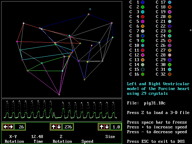 |
This is an example of 3-D data reconstruction. By measuring every distance between every crystal, the three-dimensional relationship of all the crystals can be mathematically determined, resulting in an X,Y,Z location in space for each crystal. By using our 3-D data viewing software, the user can render the resulting 3-D data by adding connecting lines between crystals and adjusting the viewing angle and playback speed. In this image you can see the left and right ventricles as defined by 29 crystals placed on the epicardium and the septal wall. |
Return to Sonometrics Home Page |